

Meet the people behind this publication


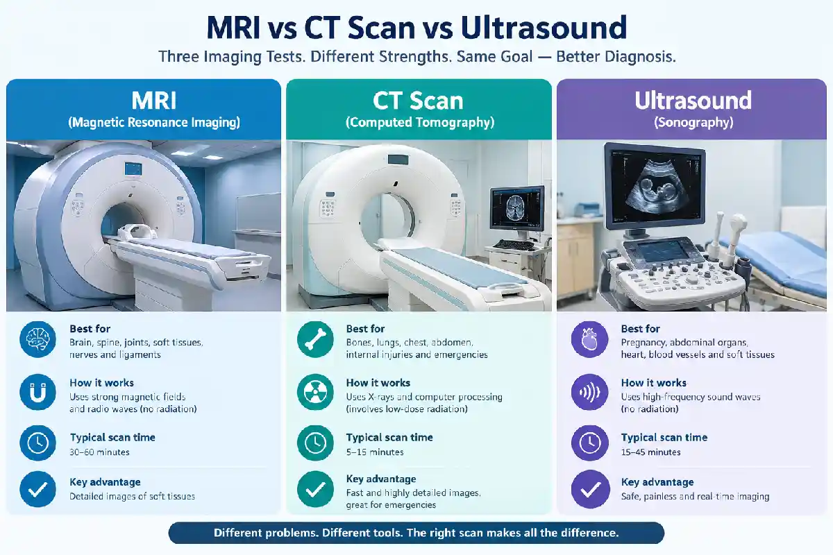
MRI vs CT Scan vs Ultrasound: When a doctor needs to see inside the body, they may use MRI, CT or ultrasound. All three create pictures of internal structures without surgery, but they use different technology and answer different questions. A test that works well for a knee injury may tell a doctor very little about a gallbladder, and the reverse is also true.
This guide explains how each type of medical imaging works, what it is commonly used for and how doctors choose between them. It is general education, not personal medical advice, and the choice of test belongs to your doctor.
This article belongs in Health TechnologyMagnetic resonance imaging (MRI) uses a strong magnetic field, radio waves and a computer to produce detailed cross-sectional images. It does not use ionizing radiation, the type involved in X-rays and CT scans.
MRI is especially useful for soft tissue, which includes the brain, muscles, ligaments and many internal organs. Doctors commonly use an MRI scan to examine the brain, spine, joints such as the knee and shoulder, the abdomen and pelvis, and the heart and blood vessels.
Most MRI exams take 15 to 45 minutes, and some run an hour or longer. You lie on a padded table that slides into a large, tube-shaped scanner and need to stay still. The machine makes loud tapping and knocking sounds, so staff provide earplugs or headphones. If enclosed spaces make you uneasy, mention it beforehand. Depending on the facility, options may include a mild sedative, a companion in the room or a more open scanner.
Safety screening matters more for MRI than for the other two tests. The magnet can pull on certain metal objects and interfere with some implanted devices. You will be asked about pacemakers, surgical clips, medication pumps, cochlear implants and metal fragments. Many newer implanted devices are MR Conditional, meaning MRI may be possible under specific conditions, but the imaging team needs the exact device information first.
A computed tomography (CT) scan takes X-ray measurements from many angles and uses a computer to build cross-sectional images. Unlike MRI, CT does involve ionizing radiation, and the dose varies by scan type. Radiology societies note there is no conclusive evidence that the small doses used in CT cause cancer, but because a very slight risk cannot be ruled out, a CT is recommended when the expected benefit outweighs it. Children are more sensitive to radiation, so pediatric scans use low-dose techniques and are done only when needed.
CT is widely used for injuries after trauma, chest and abdominal conditions, fractures, internal bleeding, appendicitis, blood clots in the lungs and the detection and monitoring of many cancers. It shows bone, soft tissue and blood vessels in the same scan.
Speed is a major reason CT is chosen. The NHS says a CT appointment usually takes 10 to 20 minutes, and in emergencies the images can reveal internal injuries and bleeding quickly. CT is also less affected by movement than MRI, and an implanted medical device will not rule it out the way it can for MRI. For the brain, joints and internal pelvic organs, however, MRI can often show soft tissue more clearly.
Ultrasound, also called sonography, uses high-frequency sound waves. A technician moves a handheld device called a transducer over gel on the skin, and a computer turns the returning echoes into images. Ultrasound does not use ionizing radiation, and standard diagnostic ultrasound has no known harmful effects.
Common uses include monitoring pregnancy and the baby, and examining the liver, gallbladder, kidneys, thyroid, breasts and blood vessels. Doppler ultrasound shows blood flow, which can help detect clots or narrowed vessels. It is also used to guide needle biopsies.
Images appear in real time, so a doctor can watch movement such as blood flow or a beating heart. The equipment is widely available, can be portable and often needs little preparation.
Sound waves are disrupted by air and gas, so ultrasound is less useful for air-filled bowel or lungs. It also cannot pass through bone, so it shows only the outer surface of bony structures.
| MRI | CT scan | Ultrasound | |
| Technology | Magnetic field and radio waves | X-rays and computer processing | High-frequency sound waves |
| Radiation | No ionizing radiation | Yes, ionizing radiation | No ionizing radiation |
| Best suited for | Brain, spine, joints, soft tissue, certain organs | Trauma, chest, abdomen, bleeding, fractures, emergencies | Pregnancy, abdominal organs, thyroid, blood vessels, guiding biopsies |
| Speed | Slower, often 15 to 45 minutes or more | Images are acquired quickly; the full appointment takes longer | Real-time images; time depends on the area examined |
| Soft-tissue detail | Excellent | Good, with bone and vessels in the same scan | Good for some organs, limited by depth, bone and air |
| Major limitations | Long, noisy and enclosed; implant screening needed | Uses radiation; less soft-tissue detail than MRI in some areas | Cannot see through bone or air-filled structures |
In hospitals, these technologies are usually run from a radiology department, where radiologists (doctors who specialize in reading medical images) review the results and report to the referring doctor. Samitivej Hospital in Bangkok, for example, lists MRI, CT and ultrasound among its diagnostic imaging services, alongside X-ray and mammography.
There is no single best scan. Doctors weigh several factors:
Discuss the options with your doctor, who can recommend the test most likely to answer the clinical question. The treating physician has your history, examination findings and earlier results. It is reasonable to ask why a test was recommended and what it is expected to show.
Contrast agents help certain tissues and blood vessels stand out more clearly on medical images. They temporarily change how those structures appear, and most of the contrast material is subsequently cleared from the body.
CT usually relies on iodine-based (iodinated) contrast, injected into a vein or taken by mouth. MRI uses gadolinium-based contrast, injected into a vein. Not every scan requires contrast, and your doctor or radiologist decides based on what needs to be seen. They also consider your medical history, any allergies and, where relevant, your kidney function.
Mild effects such as a warm flush, a metallic taste or nausea are the most common. Serious reactions are very uncommon, and radiology departments are equipped to treat them. Gadolinium is less likely to cause an allergic reaction than iodine-based contrast.
Your imaging team will give specific instructions, but some points apply broadly:
Some situations call for prompt imaging, including a serious head injury, a suspected stroke, severe abdominal pain, major trauma and possible internal bleeding. Doctors often choose a fast test such as CT in these cases, but that is not true of every emergency. Depending on the condition and the findings so far, a doctor may use CT, ultrasound, MRI or a combination. If you or someone near you has symptoms like these, seek emergency care right away rather than waiting for a scheduled scan.
Neither is better overall. MRI offers more detail for soft tissues such as the brain, spine and joints, while CT is faster and excels at showing bone, bleeding and injuries. The better test is the one that answers your doctor’s specific question.
MRI avoids ionizing radiation, which is an advantage for some patients, but it has its own safety considerations around metal implants and devices. CT uses ionizing radiation, with the dose depending on the examination, but it is unaffected by most implants and is much quicker. Which is safer depends on the individual situation.
Sometimes. For pregnancy, the gallbladder and the thyroid, ultrasound is often the first and only test needed. But sound waves cannot pass through bone or air, so a doctor may follow an ultrasound with MRI or CT when a deeper or clearer view is needed.
The two tests have different strengths. CT can quickly reveal bleeding, fractures or the general extent of a problem, while MRI can add fine soft-tissue detail. Using both can give a more complete picture.
A radiologist reviews the images and sends a report to the doctor who ordered the scan, who then explains the findings. Timing varies by facility, scan type and urgency. Urgent scans are reviewed quickly, while routine reports can take longer.

Syed Sadiq is a health education content researcher who writes evidence-informed guides on symptoms awareness, preventive health habits, and digital safety topics affecting students and families. His articles focus on helping readers understand health-related information clearly before making decisions. Through DionHealthWeb, he publishes structured health-awareness content designed to support informed conversations between readers and healthcare professionals.